Ultrasound technology stands out as a cornerstone of medical imaging, offering a cost-effective solution compared to more complex modalities, precision, ease-of-use and comfort for the patient.
While the medical ultrasound transducer market is structured with mature piezoelectric sensors, it keeps evolving with innovations driven by AI, compactness, speed, ease-of-use and accuracy. On the industrial side, trends in the semiconductor are also enabling breakthroughs with economy of scale, miniaturization and mass production of MEMS (CMUT/PMUT) transducers.
Theses evolutions allow for very positive forecast in the medical imaging market with a global ultrasound imaging systems market expected to grow steadily to a USD 3.4B revenue in 2025 (6.7% CAGR 2019-2025) according to Yole Group.
In this paper, you will learn about advances at the crossroads of two major drivers of the 2024 ultrasound medical imaging landscape and the pivotal role played by ASICs supporting these trends.
Miniaturization of ultrasound systems
When we talk about ultrasound medical imaging, there is one major physical rule: image quality degrades with the organ depth. Our skin, and other soft tissues are natural sound attenuators and can hide deep structures. If ones want to overcome these obstacles, one must either increase the power, wavelength or number of sensors or get passed them and “look” behind. The first solution is straightforward but will soon hit safety regulations in a losing competition with other medical imaging solutions like X-ray or MRI.
Until recently, the second solution to “look behind” was complex and interfered too dramatically with a major principle which is to limit patient discomfort and risks for his own safety. This is why miniaturization is a major strategy for ultrasound medical imaging today.
Ultrasound catheters or transesophageal probes, for example, are applications that clearly demand for miniaturization, allowing increasingly better functionalities in a limited form factor. Being among the least invasive solutions in the field of interventional cardiology confer upon them a strong potential for further developments.
Miniaturization is key for cutting-edge applications, such as ultrasound patches, as its feasibility relies on the development of advanced integration technics. However, more conventional applications, such as transthoracic probes, strongly benefit also from miniaturization trends.
2D arrays & 3D imaging
In parallel, innovations such as AI, transducer improvement, form factor or architecture evolutions led to major advancements in 3D imaging.
This technology presents several advantages: improved visualization, enhanced diagnostic accuracy, or better treatment planning. Overall, 3D imaging enhances the capabilities of medical imaging by providing a more detailed and accurate representation of anatomical structures, which in turn improves patient care and outcomes.
If 3D imaging represents a great interest, it also implies integrating a 2D transducer matrix into a probe thus an increased difficulty of integration in comparison to 1-D transducers. The addition of a dimension indeed involves an increase in the number of elements and thus the number of signals to manage, which complicates the interconnection between the probe and the system for a 2D matrix or a reconfigurable 2D array.
As it is not 3D imaging itself but the number of elements to manage that leads to probe development challenges, although 1.5D or 1.75D transducer probes are intended for 2D imaging, their development imply similar technics.
This is at the intersection of the needs of miniaturization and of 2D transducer matrices that arises the solutions of active probes.
Passive Vs Active probes
Passive ultrasound probes
In conventional ultrasound medical imaging systems, the catheter introduced into the heart is entirely passive. The ultrasound transducers integrated into it are generally made from a piezoelectric material, but it can alternatively be based on micro-electro-mechanical systems (MEMS) such as CMUT or PMUT.

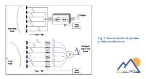
As depicted in Figure 1, each transducer element of the probe is connected to the system via an individual micro-coaxial cable part of a probe cable bundle. A switch (T/R switch) enables the connection of these elements to either the transmission chain (TX) to excite the transducers with relative delays and generate ultrasonic waves in the medium at a desired transmission focal point, or to the reception chain (RX) to record the sum of returning echoes on the elements each delayed according to a separate receiving focal point. During transmission, the system produces signals in the megahertz range or tens of megahertz, within the bandwidth of the transducer. The transmit signal amplitudes are on the order of a few tens of volts to provide sufficient energy into the medium and to ensure an exploitable signal-to-noise ratio.
The major limitation of passive probes is that each element must be connected individually to the system, which complicates integration using very thin cables, especially for transesophageal and intracardiac probes. Additionally, thinner connecting cables lead to increased attenuation during reception.
This limitation prevents the transition to 3D probes since moving to 2D transducer arrays implies a significant increase in the number of elements, limited by integration constraints.
Active ultrasound probes
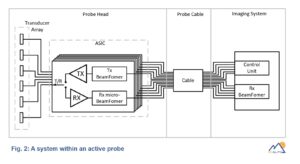
The implementation of an ASIC in the probe head enables the realization of real-time 3D probes. Part of the ultrasound scanner electronics is transferred to the ASIC, significantly reducing the number of signals to be transferred from the probe head to the system and conditioning them to limit cable-related attenuation.
For this purpose, the ASIC must integrate pulse generators and the transmit beamformer to excite the elements of the transducer, typically by switching voltages provided by the system. The ASIC reception chain must include a low-noise amplifier (LNA), a variable gain stage acting as a time-gain compensation (TGC) amplifier to compensate for signal attenuation along the ultrasonic wave path, a cable count reduction solution and one or more drivers for signal conditioning. The functionality of the T/R switch must also be ensured by the ASIC.

The reduction in the number of cables starts with pulse transmission. This is achieved by the ASIC, which generates all necessary signals based on the beamforming configured by the system. The cables connecting each element to a passive probe configuration are replaced in transmission by fewer connections.
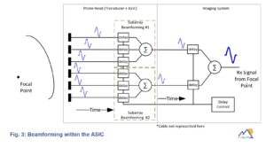
In reception, one of the most powerful signal reduction tools is micro-beamforming. This technique involves dividing the transducer into groups of elements and and to perform operation on those groups of elements instead of on single elements, as illustrated in Figure 3, generating a single signal per group. The signal from micro-beamforming is transmitted to the system instead of those directly from the elements, allowing for a reduction factor equal to the number of elements per group.

The benefits of using ASIC in ultrasonography medical imaging systems
The use of Application-Specific Integrated Circuits (ASICs) in medical imaging has been a significant trend in recent years, driven by several factors:
- Miniaturization and integration: ASICs allow for the integration of complex functionalities into smaller and more compact devices. In medical imaging, this has led to the development of more portable and lightweight equipment, enhancing mobility and accessibility.
- High performances: ASICs are specifically designed to meet the demands of medical imaging applications, offering enhanced performance in terms of speed, accuracy, and power efficiency. This enables higher-resolution imaging, faster processing speeds, and improved overall image quality.
- Customization and optimization: ASICs can be tailored to the specific requirements of different imaging modalities and applications, allowing for optimization of performance parameters such as signal processing algorithms, image reconstruction techniques, and data transmission protocols. Custom form factor is another important benefit.
- Integration with other technologies: ASICs can be seamlessly integrated with other advanced technologies such as sensors, actuators, and communication interfaces, enabling the development of multifunctional imaging systems with enhanced capabilities.
- Innovations in imaging techniques: As new imaging techniques and modalities emerge, such as 3D imaging, spectral imaging, and functional imaging, an ASIC is the solution to support these advanced functionalities.
Overall, the trend towards the use of ASICs in medical imaging reflects a growing emphasis on innovation, efficiency, and security in healthcare domain.

Ultrasound imaging innovation is facing challenges in which our ASIC design expertise is key. On the other hand, a deep understanding of the challenges involved in our customer's applications is a must. ASIC design for innovative technologies implies strong collaboration. We at IC'ALPS have a strong focus on developing ASICs for innovative medical applications. This focus, together with our ISO 13485:2016 certified development process, makes the difference in addressing our customer's needs. We are committed to contributing to the innovation effort that is leading to the Healthcare technology of tomorrow.
Jon Iriarte Martin
ASIC Application Engineer

